3D cell culture of the liver
Papers selected and presented by PhD student Jonathan Temple (he will officially become Editor once he has published his first article).
Editor Raphaël Lévy
Paper supported long-term 3D liver co-culture model for the assessment of hepatotoxic drugs (2018)
Yaqing Wang, Wentao Su, Li Wang, Lei Jiang, Yang Liu, Lijian Hui, Jianhua Qin
PubMed: 30090558 DOI: 10.1039/c7tx00209b
Paper is an attractive biocompatible material for cell-based applications due to its biocompatibility. In this study, Wang et al. established a simple paper-based scaffold array for creating a 3D liver co-culture model that enabled the assessment of drug induced hepatotoxicity. Using hiHeps, generated from fibroblasts by lineage conversion, co-cultured with HUVECs Wang et al. demonstrate the ability of the paper scaffold to maintain liver specific functions of producing albumin and urea for up to 2 months (figure 5 in Wang et al). In addition, the hiHeps in this co-cultured model maintained a higher expression of cytochrome P450 genes as compared with a monolayer culture on a plate and a single culture on paper of hiHeps, revealing a marked enhancement of hepatic functions in the 3D liver co-culture model (figure 6 in Wang et al).
This paper demonstrates well the possibility of using filter paper as a substrate for cell culture, but this might be more appropriately described as sandwich culture than a true 3D culture, as illustrated in the schematic (figure 1 of Wang et al) and confirmed by the SEM and fluorescence imaging (Figure 1).
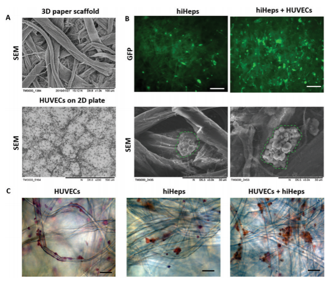
Figure 1. SEM and fluorescence imaging of hiHeps on the filter paper scaffold (reproduced from Figure 2 of Wang et al)
It appears that the cells are growing in cell clumps (spheroids) on top of the filter paper and not actually utilising the scaffold material itself.
My second main issue with the paper is that they, like many others, do not count the number of cells that stick to the scaffold. Wang et al. even mention they believe cells detach from the scaffold during the culture however, they still normalise to the number of cells seeded. This is less of an issue with this model compared with other papers as all the cells should still remain in the well however, not all will be growing in 3D.
The paper also makes a lot of claims such as; they identified cell morphology, the cells had uniform distribution, HUVECS grew along the axis of the scaffold, the cells assembled properly, 3D liver tissue displayed a normal morphology, suggesting the migration and reassembly of the two cells during the course of the co-culture without backing up any of the statements with data or if the data was shown the conclusions drawn from it were without sufficient evidence.
Overall, I like the idea behind the culture and Wang et al. do demonstrate that the filter paper culture does improve hiHep function but the paper makes a lot of assumptions without backing them up and would benefit from a more in-depth analysis.
-(2).jpg)