3D cell culture of the liver
Papers selected and presented by PhD student Jonathan Temple (he will officially become Editor once he has published his first article).
Editor Raphaël Lévy
Development of a 3D cell printed construct considering angiogenesis for liver tissue engineering (2016)
Jin Woo Lee, Yeong-Jin Choi, Woon-Jae Yong, Falguni Pati, Jin-Hyung Shim, Kyung Shin Kang, In-Hye Kang, Jaesung Park, Dong-Woo Cho
PubMed: 26756962 DOI: 10.1088/1758-5090/8/1/015007
This article demonstrates a novel 3D cell printing technology and its application in liver tissue engineering. Currently the poor mechanical properties of cell-laden hydrogels cause issues during the printing process. Using polycaprolactone (PCL) as a framework material Lee et al (2016) demonstrate how they were able to use a multi-head tissue/organ building system (MtoBS) to produce a printed, 3D model of the liver. The PCL framework has ‘excellent mechanical properties’ which supports the cell containing hydrogel from either side (figure 1).
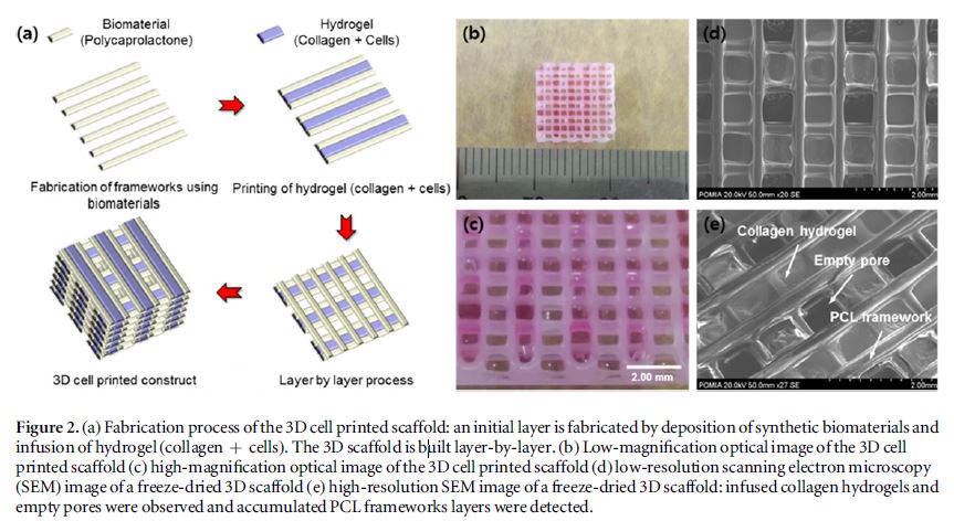
Figure 1. Reproduced from Lee et al (Figure 2); fabrication process of the 3D printed construct
Lee et al (2016) compared three different groups of cells; Group one included only primary hepatocytes (HC), group two included a 1:3 mixture of HCs and human lung fibroblasts (HLF), and group three contained a 1:3:1 mixture of HCs, HLFs and human umbilical vein endothelial cells (HUVEC) printed into the 3D model. They performed metabolic activity assays (Albumin and Urea) along with confocal microscopic analysis to study the models. They found much higher metabolic activity for group three than the other two groups and attributed this to the interaction between the different cell types as they release various transcription factors which lead to an increase in urea and albumin expression (figure 2). They also showed confocal images or albumin staining that further supported an increase in albumin production in group three.
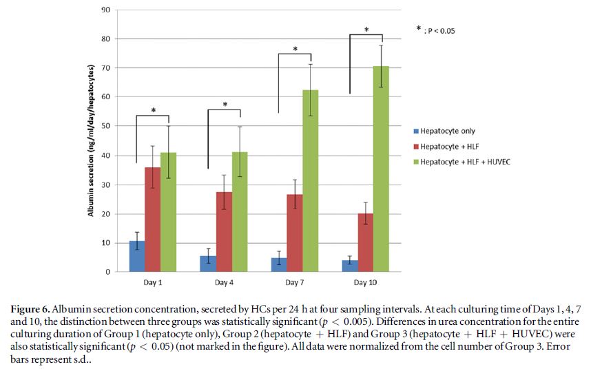
Figure 2. Reproduced from Lee et al (Figure 6); albumin production across the three groups. Group three has significantly higher albumin production and is also the only group in which production increases over time.
This paper demonstrates well the ability to efficiently print large-volume 3D constructs that have a stable micro-environment for liver tissue engineering. They however, have performed little analysis on the model other than simple biochemical readouts along with confocal imaging which shows very little other than cell growth over a 10 day period.
My main issue with this paper is that they claim that the model has ‘capillary-like formation’ as well as ‘vascularization of the construct’. They show no evidence for this other than the presence and growth of HUVEC cells. Their hypothesis that the increased urea and albumin production is due to the release of soluble factors between the cell types is reasonable but they do not show convincing evidence that metabolic increase can be attributed to ‘vascularisation’.
Some important experimental information seems to be missing, in particular: 1) the stain visualised in figure 4 is not specified; 2) most data are normalized by numbers of cells but it is unclear on which day and how this was measured.
Overall, I like the concept of this paper and I think that the printing technique that they use is novel and overcomes the problems faced when printing hydrogels with poor mechanical properties.
-(2).jpg)